The development of X-ray technology has been a cornerstone of medical diagnostics, allowing physicians to observe the internal structures of the human body non-invasively. At the forefront of this technological advancement is the X-ray flat panel detector. This article explores the functionality, benefits, and applications of X-ray flat panel detectors, highlighting their transformative impact on medical imaging.

Understanding The X-Ray Flat Panel Detector
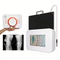
An X-ray flat panel detector (FPD) is an advanced digital imaging device that converts X-ray radiation into detailed, high-quality digital images. The device consists of two primary components: the detector panel and the control unit.
1. The Detector Panel
The detector panel is a sophisticated component, usually constructed from scintillator materials such as cesium iodide (CsI) or gadolinium oxysulfide (Gd2O2S). These materials are pivotal because they convert X-ray photons into visible light photons. Here's how it works:
Scintillation: When X-rays pass through a patient and hit the scintillator layer of the detector panel, the scintillator absorbs the X-ray photons and re-emits them as light.
Light Detection: The emitted light is then captured by a layer of photodiodes underneath the scintillator, which convert the light into electrical signals. This conversion is essential for digitizing the X-ray information.
2. The Control Unit
The control unit processes the electrical signals from the detector panel. It manages the following:
Signal Conversion: The control unit converts the analog electrical signals into digital data.
Image Processing: It applies algorithms to enhance the image quality, adjusting parameters such as contrast and sharpness.
Parameter Modulation: The control unit also allows for real-time modulation of X-ray exposure settings, including dosage and imaging parameters, to optimize the image quality and minimize patient exposure to radiation.
Benefits Of X-Ray Flat Panel Detectors
X-ray flat panel detectors offer numerous advantages over traditional X-ray film technology and computed radiography (CR) systems.
1. Enhanced Image Quality
FPDs provide superior image quality with higher spatial resolution and better contrast compared to film and CR systems. This improvement is crucial for accurate diagnosis as it enables the visualization of finer anatomical details and subtle pathological changes.
2. Reduced Radiation Exposure
The efficiency of FPDs allows for lower radiation doses to achieve high-quality images. Since images can be reviewed in real-time, adjustments can be made promptly, reducing the need for repeat exposures and thus lowering the cumulative radiation dose for patients.
3. Faster Imaging Process
FPDs streamline the imaging process by eliminating the need for film development or image processing in CR systems. The immediate availability of digital images facilitates quicker diagnosis and treatment decisions, improving patient throughput and operational efficiency in medical facilities.
4. Digital Integration and Storage
The digital nature of FPDs simplifies the integration with Picture Archiving and Communication Systems (PACS). Digital images can be easily stored, retrieved, and shared among healthcare providers, enhancing collaborative care and long-term patient record management.
Utilization In Medical Imaging
X-ray flat panel detectors are employed in a wide range of medical imaging applications, reflecting their versatility and effectiveness.
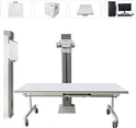
1. Radiography
In general radiography, FPDs are used for capturing detailed images of various body parts, including the chest, abdomen, and extremities. They are particularly valuable in emergency settings where rapid image acquisition is essential for prompt diagnosis and treatment.
2. Fluoroscopy
FPDs are integral to fluoroscopic procedures, which provide real-time imaging of the body's internal functions. They are used in gastrointestinal studies, angiography, and interventional radiology, where live imaging guides surgical procedures and therapeutic interventions.
3. Computed Tomography (CT) Scans
Advanced FPDs are utilized in some CT systems, offering improved image quality and faster scanning times. Their high resolution and sensitivity enhance the detection of abnormalities in various organs and tissues.
4. Orthopedic Imaging
In orthopedic imaging, FPDs deliver high-resolution images of bones, joints, and soft tissues. This capability is crucial for diagnosing fractures, joint dislocations, and degenerative conditions, enabling precise surgical planning and monitoring of treatment outcomes.
5. Dental Imaging
FPDs are widely used in dental radiography, providing detailed images of teeth, gums, and jaw structures. They are essential for diagnosing dental issues, planning treatments, and conducting routine dental check-ups with minimal radiation exposure.
6. Mammography
In mammography, FPDs play a critical role in the early detection of breast cancer. They provide high-quality images that can reveal small tumors and microcalcifications, crucial for early intervention and improved patient outcomes.
Technological Advancements and Future Prospects
The ongoing advancements in X-ray flat panel detector technology promise to further revolutionize medical imaging. Key areas of development include:
1. Improved Sensitivity and Resolution
Research is focused on enhancing the sensitivity and spatial resolution of FPDs. Advances in scintillator materials and photodiode technology are expected to produce even finer image details, improving diagnostic accuracy.
2. Dose Reduction Techniques
Innovations in dose reduction techniques aim to minimize patient exposure while maintaining or enhancing image quality. These include the development of algorithms for image reconstruction and noise reduction, allowing for lower radiation doses without compromising diagnostic capabilities.
3. Integration with Artificial Intelligence (AI)
The integration of AI with FPDs holds significant potential for automating image analysis and interpretation. AI algorithms can assist in detecting abnormalities, enhancing image quality, and streamlining workflows, thereby increasing the efficiency and accuracy of radiological diagnostics.
4. Portability and Versatility
Future FPD designs may focus on increased portability and versatility, enabling their use in diverse clinical settings, including remote and resource-limited environments. This development will make high-quality imaging accessible to a broader range of patients.
Conclusion
X-ray flat panel detectors represent a transformative advancement in medical imaging, providing superior image quality, reduced radiation exposure, and streamlined imaging processes. Their extensive applications across various medical disciplines underscore their versatility and efficacy. As technological advancements continue to evolve, X-ray flat panel detectors are set to further enhance diagnostic imaging, offering new possibilities for improved patient care and outcomes.
The future of medical imaging lies in the continued innovation and integration of FPD technology, ensuring that healthcare professionals can deliver precise, efficient, and safe diagnostic services. The revolution in X-ray technology driven by flat panel detectors marks a significant leap forward in the quest for better healthcare solutions, ultimately benefiting patients and healthcare systems worldwide.