In the realm of medical diagnostics, X-ray technology stands as a cornerstone in identifying and treating a myriad of conditions. The development and diversification of X-ray imaging equipment have significantly enhanced the ability of healthcare professionals to examine different regions of the body, leading to more accurate diagnoses and effective treatments. This comprehensive guide provides an overview of the various types of X-ray equipment utilized in today's healthcare facilities, emphasizing their unique functions and applications.
1. CT Scanners (Computed Tomography)
CT scanners are the pinnacle of modern medical imaging, utilizing X-ray technology to produce detailed cross-sectional images of the body. These devices emit X-rays from multiple angles around the patient and use a computer to process the data, creating either a two-dimensional slice or a three-dimensional rendering of the scanned area. The ability to generate highly detailed images makes CT scanners invaluable in detecting even minor abnormalities, such as small tumors or vascular anomalies. Their applications extend across various medical fields, including oncology, cardiology, and neurology.
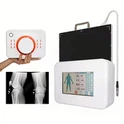
2. X-Ray Photography Devices
X-ray photography devices are essential for capturing two-dimensional images of the body's internal structures. These devices work by emitting X-rays that pass through the patient and are absorbed at different rates by various tissues. The resulting images are detected and captured on digital radiography (DR) systems, computed radiography (CR) systems, or traditional screen-film. DR systems offer real-time image display and superior image quality, while CR systems provide a balance of digital capabilities and cost-effectiveness. Traditional screen-film remains in use in some settings due to its simplicity and reliability.
3. Gastrointestinal X-Ray Machines
Gastrointestinal X-ray machines are specialized for imaging the gastrointestinal tract. These fluoroscopic devices allow for dynamic imaging and real-time observation of the digestive system during procedures like barium swallows and small bowel follow-through. They are also employed for diagnostic and interventional radiographic procedures, such as identifying blockages, ulcers, and other abnormalities within the GI tract. Their ability to provide continuous imaging is crucial for accurate diagnosis and treatment planning.
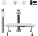
4. General X-Ray Machines
General X-ray machines, also known as fluoroscopy units, are versatile devices used to visualize a wide range of internal structures. These machines work by emitting X-rays that penetrate different materials, with varying absorption rates depending on tissue type. This variation in absorption allows for the creation of detailed images that are critical for diagnosing conditions like fractures, infections, and tumors. Fluoroscopy units are widely used in both diagnostic and interventional radiology.
5. Mammography Machines
Mammography machines are specialized X-ray devices designed specifically for imaging breast tissue. They play a crucial role in the early detection of breast cancer by providing high-resolution images that can reveal tumors and other abnormalities in breast tissue. Mammography is a key component of women's healthcare, with routine screenings recommended for early diagnosis and better outcomes. Advanced digital mammography systems offer enhanced image quality and reduced radiation exposure compared to traditional film-based systems.
6. DSA (Digital Subtraction Angiography)
Digital Subtraction Angiography (DSA) units represent high-end imaging technology essential for interventional radiology. These systems are particularly useful for visualizing blood vessels and conducting minimally invasive vascular procedures. DSA works by capturing images before and after contrast injection and then subtracting the pre-contrast image from the post-contrast image, highlighting vascular structures with exceptional clarity. This technique is instrumental in diagnosing and treating conditions such as aneurysms, arterial blockages, and venous malformations.
7. Mobile X-Ray Units
Mobile X-ray units are designed for flexibility and portability, allowing for on-site diagnostic imaging across various departments. These units are mounted on wheels, enabling easy transport to different locations within a healthcare facility. Mobile X-ray units come in various forms, including mobile C-arm, digital radiography (DR), computed radiography (CR), and traditional screen-film units. Their portability makes them ideal for use in emergency departments, intensive care units, and during bedside examinations where moving the patient is not feasible.
8. Lithotripters (ESWL - Extracorporeal Shock Wave Lithotripsy)
Lithotripters are specialized devices used in the non-invasive treatment of kidney stones and other calculi. Extracorporeal Shock Wave Lithotripsy (ESWL) uses focused shock waves to fragment stones into smaller pieces that can be naturally expelled by the body. An integrated X-ray unit is often used to accurately locate the stones before treatment. This method is preferred for its minimal invasiveness and high success rate in treating urolithiasis.
9. Molecular Imaging (SPECT/CT and PET/CT)
Molecular imaging combines nuclear medicine techniques with computed tomography (CT) to provide detailed images of both the anatomical and functional aspects of the body. Single Photon Emission Computed Tomography (SPECT) and Positron Emission Tomography (PET) detect gamma radiation emitted by the body after the administration of a radiotracer. When fused with CT imaging, these modalities offer precise localization and characterization of diseases, making them indispensable in oncology, cardiology, and neurology for diagnosing conditions such as cancer, heart disease, and neurological disorders.
10. Dental X-Ray Machines
Dental X-ray machines are crucial for dental diagnostics and treatment planning. These devices include intraoral film units for capturing detailed images of individual teeth and panoramic units for broader views of the entire mouth. Advanced dental imaging systems also include oral CT scanners, which provide three-dimensional images for complex dental procedures such as implants and orthodontic assessments. Modern dental X-ray machines offer enhanced image quality and lower radiation doses, improving patient safety and diagnostic accuracy.
11. Linear Accelerators
Linear accelerators (linacs) are used in radiation therapy to treat cancer. These sophisticated machines generate high-energy particles, such as electrons, protons, or heavy ions, which are directed at tumors to destroy or reduce their size. Linear accelerators are capable of delivering precise doses of radiation to specific areas, minimizing damage to surrounding healthy tissues. They are a cornerstone of modern radiotherapy, used in the treatment of various cancers, including breast, prostate, and brain tumors.
Conclusion
X-ray imaging equipment forms the backbone of modern medical diagnostics, providing critical insights into the human body that facilitate accurate diagnoses and effective treatments. Each type of X-ray machine serves a distinct purpose, from general radiography and specialized mammography to advanced molecular imaging and therapeutic applications. Understanding the specific functions and applications of these machines is essential for healthcare providers aiming to deliver optimal patient care. For facilities seeking to upgrade or acquire new X-ray equipment, collaborating with reputable manufacturers and staying informed about technological advancements is crucial. By selecting the appropriate X-ray machine tailored to their needs, healthcare facilities can enhance their diagnostic capabilities, improve patient outcomes, and ensure a high standard of care.
For more information on X-ray technology or to explore equipment options, consult with professional entities and manufacturers who specialize in medical imaging solutions. Stay updated on the latest advancements to keep your facility at the forefront of medical diagnostics.