
types of ultrasound probes and their uses
Ultrasound probes are vital tools in medical imaging. They convert electrical energy into sound waves. These sound waves create images of the body's internal structures.
Different types of ultrasound probes serve various medical needs. Each probe type is designed for specific anatomical areas. This ensures accurate and detailed imaging.
Linear probes are perfect for vascular imaging. They are also used for small parts like the thyroid. Their high frequency provides excellent resolution.
Curvilinear probes are ideal for abdominal scans. They offer a wider field of view. This makes them suitable for obstetric imaging.
Phased array probes excel in cardiac imaging. They can capture heart images through the ribs. This is crucial for diagnosing heart conditions.
Endocavitary probes are used for internal exams. They are essential for transvaginal and transrectal imaging. These probes provide detailed internal views.
Understanding ultrasound probe types is crucial. It helps medical professionals choose the right tool. This ensures accurate diagnosis and effective treatment planning.
What is an Ultrasound Probe? Understanding the Basics
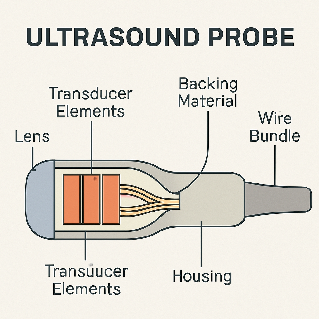
An ultrasound probe is a critical component in medical diagnostics. It's a handheld device that emits sound waves. These waves penetrate the body to produce images of internal structures.
The functioning of these probes rests on the piezoelectric effect. This effect involves converting electrical energy into mechanical vibrations. The probe sends and receives these sound waves.
Ultrasound probes are also known as transducers. They play a significant role in non-invasive imaging. This makes them a preferred choice for various medical exams.
There are various ultrasound probe types, each with unique features. These differences cater to specific anatomical regions and imaging needs. Some key features include frequency, shape, and field of view.
Understanding these basics helps in selecting the right tool for the job. These choices directly impact the quality of diagnostic images. Knowledge of the types of ultrasound transducer probes allows for optimized patient care.
How Do Ultrasound Probes Work?
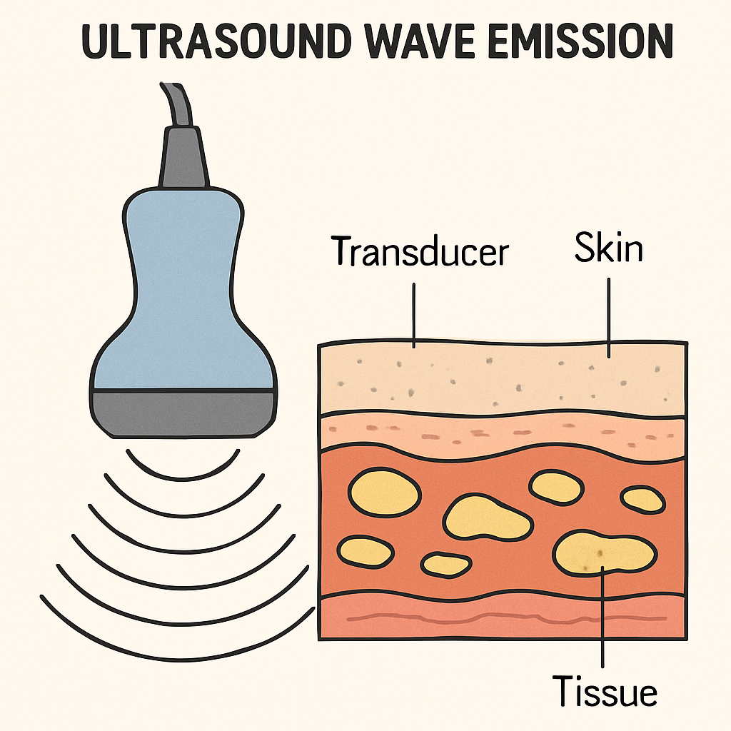
Ultrasound probes operate based on sound wave principles. They emit high-frequency sound waves through the body, which then reflect back. This reflection creates a real-time image on the monitor.
The process involves several key steps. First, the ultrasound machine sends electrical currents to the probe. This current generates sound waves.
The Main Functions of Ultrasound Probes Include:
Emitting Sound Waves: The probe sends sound waves into the body.
Receiving Echoes: Captures the returning sound waves after they bounce back.
Processing Data: Converts these sound echoes into visible images.
The probe's frequency influences image clarity and depth. Higher frequencies provide sharper images but penetrate less deeply. Lower frequencies reach deeper but offer less detail.
These tools are versatile and can adapt to different medical needs. However, choosing the appropriate probe is crucial for effective diagnosis.
Key Factors in Choosing an Ultrasound Probe
Selecting the right ultrasound probe involves several considerations. The application's specificity is paramount. Each probe serves unique diagnostic purposes.
The frequency of the probe plays a crucial role. Higher frequencies are ideal for superficial structures, while lower frequencies suit deeper tissue imaging.
Important Considerations When Choosing a Probe:
Purpose: Consider the specific clinical application.
Frequency: Choose based on desired image detail and depth.
Compatibility: Ensure the probe fits with existing ultrasound systems.
Another factor is the probe's design. The shape can impact the ability to access certain body areas. Linear probes, for instance, are great for small parts.
Comfort and usability also matter. Probes should be ergonomic, reducing fatigue during prolonged use. Understanding these factors aids in effective decision-making.

Overview of Ultrasound Probe Types
Ultrasound probes come in various types, each designed for specific uses. Knowing these differences is essential for accurate diagnostics. Linear, curvilinear, phased array, and more contribute to diverse medical imaging needs.
Main Types of Ultrasound Probes:
Linear Probes: High frequency for surface imaging.
Curvilinear Probes: Broad view, perfect for deeper imaging.
Phased Array Probes: Ideal for cardiac imaging.
Each type serves distinct anatomical targets. Linear probes excel in detailed imaging of small parts. Curvilinear probes provide a wider field for abdomen assessments. Phased arrays deliver precise heart images through ribs.
These probes differ not only in design but also in function. Their varied frequencies and shapes cater to diverse medical requirements.

Linear Ultrasound Probes
Linear ultrasound probes are renowned for their high-frequency output, making them suitable for detailed imaging. They provide excellent resolution, ideal for observing superficial structures.
These probes have a flat scanning surface. This feature suits linear probes for vascular imaging and assessing small parts such as the thyroid and breast. Due to their high frequency, they offer precision in imaging but have limited depth penetration.
In medical settings, linear probes are frequently employed in musculoskeletal examinations. They efficiently capture images of tendons, muscles, and joints, helping diagnose disorders in these areas.
Linear probes are also a staple in vascular studies. They effectively visualize veins and arteries, assisting in evaluating blood flow and detecting blockages.
A few significant applications of linear probes include:
Vascular Imaging: Precise assessment of veins and arteries.
Small Parts Imaging: Critical for examining thyroid, breast, and testicles.
Musculoskeletal: Excellent for tendon and joint evaluations.
These probes are essential for emergency and critical care settings too. Their resolution aids in conducting quick and accurate bedside assessments.
To optimize their use, users must understand the probe's frequency range and proper handling techniques. Proper maintenance ensures continued high performance in clinical settings.


Curvilinear (Convex) Ultrasound Probes
Curvilinear ultrasound probes, often termed convex probes, are known for their broad field of view. This characteristic makes them perfect for abdominal examinations.
These probes operate at a lower frequency compared to linear probes. As a result, they offer deeper tissue penetration, essential for imaging larger organs.
In obstetrics, curvilinear probes are indispensable. They allow detailed visualization of the developing fetus during pregnancy check-ups, ensuring both mother and baby's health.
Their curved transducer head facilitates access to wide anatomical areas. This makes them highly effective for general-purpose imaging tasks, including assessments of the liver and kidneys.
Key uses for curvilinear probes include:
Abdominal Imaging: Comprehensive views of organs such as the liver, spleen, and gallbladder.
Obstetric Imaging: Monitoring fetal growth and health throughout pregnancy.
Gynecological Exams: Visualizing pelvic organs for abnormalities.
The benefits of curvilinear probes extend beyond standard examinations. Their ability to provide expansive imaging makes them valuable in emergency settings.
To maximize their utility, practitioners should select the correct frequency settings. This ensures optimal depth and resolution, crucial for diagnostic clarity.
Maintaining these probes involves regular inspection for wear and proper cleaning to prevent contamination. Their delicate design requires care to ensure longevity.


Phased Array Ultrasound Probes
Phased array ultrasound probes are unique due to their compact, sector-shaped imaging capabilities. They excel at navigating small acoustic windows, making them ideal for cardiac applications.
These probes emit narrow beams that can steer electronically. This allows clinicians to gather detailed images of complex structures like the heart.
Phased array probes are frequently used in echocardiography. Their ability to capture real-time motion of the heart's chambers and valves is crucial for diagnosing cardiac conditions.
Their design facilitates imaging between ribs, which is essential for accurate cardiac imaging. This characteristic is particularly beneficial in intensive care settings.
Key applications of phased array probes include:
Echocardiography: Assessing heart function, valve issues, and blood flow.
Emergency Medicine: Rapid diagnostics in critical situations.
Thoracic Imaging: Evaluating lung and pleural conditions.
Despite their specialized use, these probes possess versatility. They are often employed in fast-paced environments where precision is critical.
Operators must be skilled in controlling probe settings. Adjustments can affect the clarity and accuracy of heart images, impacting diagnostic outcomes.
Proper care includes careful handling to prevent damage and regular cleaning. These practices ensure the longevity and reliability of the probes.


Endocavitary Ultrasound Probes
Endocavitary ultrasound probes are specifically designed for internal examinations. They provide high-resolution images of structures within body cavities.
These probes are essential tools in gynecology and urology. By offering close proximity imaging, they help clinicians obtain detailed visuals of the anatomy.
Endocavitary probes often feature a small, curved transducer. This design allows easy insertion into body orifices for optimal image acquisition.
Key applications of endocavitary probes include:
Transvaginal Ultrasound: Assessing reproductive organs and guiding fertility procedures.
Transrectal Ultrasound: Evaluating the prostate gland and guiding biopsies.
Early Pregnancy Scans: Providing detailed images of the fetus and uterus.
Their high frequency ensures excellent image clarity but limits depth penetration. This trade-off is acceptable due to the focus on nearby structures.
The use of endocavitary probes requires patient consent and comfort considerations. Proper technique and bedside manner are crucial for patient cooperation.
Sterile technique and probe covers are standard practices to maintain hygiene. Cleanliness prevents cross-contamination and infection.
Operators must ensure the probe is handled gently to avoid damage. Routine maintenance extends the functional lifespan of the equipment.


Pencil (CW Doppler) Ultrasound Probes
Pencil probes, also known as Continuous Wave (CW) Doppler probes, are specialized for measuring blood flow velocities.
These probes are not used for imaging but excel in hemodynamic assessments. Their primary application is in vascular studies.
Pencil probes are lightweight and have a simple design with two piezoelectric crystals. One continuously emits sound waves while the other receives them.
Key applications include:
Peripheral Vascular Assessment: Analyzing blood flow in limbs.
Cardiac Studies: Evaluating heart valve functions.
Surgical Monitoring: Checking blood flow during procedures.
Their high sensitivity makes them ideal for measuring high-velocity blood flows. However, their inability to generate images requires complementary imaging techniques.
This type of probe is highly valued for its precision in blood flow analysis. Its compact size also enables easy handling.

3D and 4D Ultrasound Probes
3D and 4D ultrasound probes have revolutionized medical imaging by providing volumetric and dynamic views. These advances enhance the visualization of anatomical structures, offering a more comprehensive examination than traditional 2D probes.
The difference between 3D and 4D probes lies in motion. While 3D probes capture static volumetric images, 4D probes add the dimension of time, creating real-time video imaging.
These probes are invaluable in obstetrics, allowing detailed fetal assessments. Parents benefit from seeing lifelike images of their unborn child, while clinicians gain insights into fetal development and health.
Key applications of 3D and 4D probes include:
Obstetrics: Detailed examination of fetal anatomy.
Gynecology: Evaluating uterine and ovarian structures.
Cardiology: Assessing heart structures and functions.
Technological advancements continue to improve image quality and capture speed. These enhancements support more accurate diagnostics and outcomes.

Specialty Ultrasound Probes
Specialty ultrasound probes are designed for unique and challenging imaging scenarios. These probes extend the capabilities of ultrasound technology, addressing specific clinical needs with precision.
Intravascular ultrasound (IVUS) probes, for example, are miniature devices used within blood vessels. They provide detailed images crucial for cardiovascular procedures, helping assess plaque buildup and artery walls.
Another innovative example is the intraoperative probe. These are used during surgeries to guide procedures and confirm outcomes, offering real-time imaging support in complex environments.
Specialty probes include:
IVUS Probes: Detailed imaging inside blood vessels.
Intraoperative Probes: Used during surgeries for guidance and assessment.
Endoscopic Probes: Combined with endoscopes for gastrointestinal imaging.
These specialized tools highlight the versatility of ultrasound technology. They play vital roles across a broad spectrum of medical fields, from gastroenterology to vascular surgery.
Developments in technology consistently improve the function and efficacy of specialty probes, widening their clinical applications.

Frequency, Depth, and Resolution: How Probe Design Impacts Imaging
The design of an ultrasound probe greatly impacts its imaging capabilities. Key factors include frequency, depth, and resolution. Each plays a crucial role in determining image quality and application suitability.
Higher frequency probes provide clearer images with better resolution. They excel in detailed imaging of superficial structures like tendons and muscles. However, their penetration depth is limited, making them less effective for deeper tissues.
Conversely, lower frequency probes penetrate deeper into the body. They are ideal for imaging deeper organs but sacrifice some image detail and clarity.
Understanding the balance among:
Frequency: Affects image detail and depth.
Depth: Determines how far the probe can 'see' inside.
Resolution: Influences image clarity and detail.
Selecting the right probe hinges on the area to be examined and required image quality. Matching probe specifications to clinical needs is key to obtaining accurate and useful diagnostic images.
Technological advances continue to refine the trade-offs between these factors, improving overall imaging versatility.

Common Clinical Applications for Each Probe Type
Different types of ultrasound probes each find their niche in clinical settings. They cater to specific imaging needs across various medical fields.
Linear probes are frequently used in vascular imaging. Their high resolution aids in assessing blood vessels and small organs.
Curvilinear probes are commonly employed for abdominal and obstetric imaging. They offer a wider field, ideal for visualizing larger internal structures.
Phased array probes specialize in cardiac imaging. Their narrow beam allows detailed heart imaging, even through ribs.
Endocavitary probes are suited for internal exams. They are ideal for transvaginal and transrectal procedures, offering close-up images.
Specialty probes like 3D and 4D are used in prenatal scans. They bring fetal development to life with volumetric imaging.
Key clinical applications include:
Linear: Vascular and small parts imaging
Curvilinear: Abdominal and obstetric scans
Phased array: Cardiac assessments
Endocavitary: Internal examinations
3D/4D: Fetal development imaging
These diverse applications demonstrate the versatility and specificity of ultrasound probes in medical diagnostics.

Advances in Ultrasound Probe Technology
Ultrasound technology continually evolves, enhancing imaging capabilities and diagnostic accuracy. Recent advancements have led to more precise and versatile probes.
New materials have improved probe durability and sensitivity. This allows better sound wave transmission and clearer images.
Miniaturization of components has led to more portable ultrasound devices. These can provide real-time imaging in diverse settings, from clinics to remote locations.
Integration with artificial intelligence is on the rise. AI aids in image analysis, reducing interpretation errors and improving efficiency.
Key technological advancements include:
Material innovation: Improved transmission and image clarity
Portable designs: Enhanced accessibility in varied locales
AI integration: Increased diagnostic accuracy
These innovations make ultrasound a leading choice in non-invasive imaging.

Proper Care and Maintenance of Ultrasound Probes
Proper care of ultrasound probes extends their lifespan and ensures precise imaging. Regular maintenance is crucial for reliable performance.
Clean probes thoroughly after each use. Use approved disinfectants to prevent infection. Avoid harsh chemicals that may damage surfaces.
Inspect probes regularly for signs of wear or damage. Replace worn cables and damaged components promptly to avoid compromised imaging.
Proper storage is essential. Store probes in designated cases or racks to prevent accidental drops or impact.
Key maintenance tips include:
Use approved cleaning agents
Inspect for wear and tear
Store correctly to prevent damage
Consistent care ensures probes perform optimally, providing accurate diagnostic images.

Frequently Asked Questions About Ultrasound Probe Types
Understanding ultrasound probes can spark many questions. Here, we address common queries.
What is the difference between linear and curvilinear probes? Linear probes deliver high-resolution images for superficial structures. Curvilinear probes, with a broad field of view, are ideal for deeper imaging like abdominal exams.
How do phased array probes work? Phased array probes utilize multiple small elements that fire sound waves sequentially, capturing detailed images, especially in cardiac evaluations.
Common questions include:
What are phased array probes used for?
Why are 3D probes beneficial in obstetrics?
How is probe frequency related to resolution?
Are all probes used interchangeably? Each probe has a specific purpose. It's vital to select the right type for the intended diagnostic task to ensure optimal imaging results.
Navigating probe types helps optimize diagnostic outcomes and enhance patient care.
Conclusion: Choosing the Right Ultrasound Probe for Your Needs
Selecting the appropriate ultrasound probe is crucial for accurate diagnostics. The diversity in probe types allows for tailored imaging solutions. This ensures both patient care and clinical outcomes are optimized.
Consider the specific clinical application when selecting a probe. Think about the anatomy being examined and the depth required. Each probe's design offers unique advantages.
Key factors in probe selection include:
Probe frequency
Field of view
Clinical application
Image resolution
Always balance these factors with the benefits and limitations of each probe. Adequate training and familiarity with probe types are essential for effective usage. This knowledge empowers professionals to make informed decisions, enhancing diagnostic accuracy and patient experiences. Consistent evaluation of probe performance aids in maintaining reliable imaging standards.