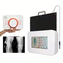
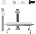
Mobile X-ray machines are indispensable in modern healthcare, providing critical diagnostic imaging services at the patient's bedside. These compact, portable devices allow healthcare professionals to perform diagnostic procedures directly in various settings, including hospitals, clinics, and even remote locations. However, the convenience and efficacy of mobile X-ray technology come with concerns about radiation exposure. This article explores the nature of radiation from mobile X-ray machines, assesses the associated risks, and provides guidelines for minimizing exposure to ensure safety for both patients and healthcare professionals.
Understanding Radiation Exposure
Radiation exposure is a critical consideration in medical imaging. Ionizing radiation, used in X-ray imaging, can penetrate body tissues to produce images of internal structures. Radiation exposure is measured in millisieverts (mSv), and the health risks associated with exposure depend on the dose. High doses of radiation can damage cellular structures and increase the risk of cancer and other health issues over time.
Radiation can come from various sources, including natural background radiation from the environment (such as cosmic rays and radon gas) and man-made sources (like medical X-ray machines). Understanding the levels of radiation exposure and the cumulative effects of repeated exposure is crucial for managing risks effectively.
Radiation Risks Associated with Mobile X-Ray Machines
Mobile X-ray machines utilize ionizing radiation to create diagnostic images. The key concern is the balance between obtaining high-quality diagnostic information and minimizing radiation exposure. Although the radiation dose from a single X-ray is relatively small, repeated or high-dose exposures can pose significant health risks.
The United States Nuclear Regulatory Commission (NRC) reports that a single chest X-ray typically delivers about 0.1 mSv of radiation. For perspective, this is roughly equivalent to the radiation exposure a person receives from natural background sources over three days. However, this cumulative exposure from multiple X-ray examinations can increase the risk of adverse health effects.
Factors Affecting Radiation Exposure
Several factors influence the radiation dose received during an X-ray examination:
Beam Intensity and Duration: The amount of radiation used to generate the X-ray image.
Distance from the Source: The closer the patient is to the X-ray source, the higher the radiation dose.
Use of Shielding: Protective measures, such as lead aprons and shields, can significantly reduce exposure.
Patient Size and Composition: Larger patients or those with dense tissue may require higher doses for clear imaging.
X-Ray Machine Settings: Optimizing the machine's settings, including voltage (kVp) and current (mA), helps control the radiation dose.
Managing Radiation Exposure: Best Practices
To ensure the safety of both patients and healthcare staff, it is essential to implement best practices for reducing radiation exposure from mobile X-ray machines:
1. Use Shielding
Lead Protection: Implement lead shielding to protect both patients and healthcare professionals. Lead aprons, thyroid shields, and lead-lined walls or doors are effective at blocking or reducing radiation exposure. Ensure that all protective equipment is properly fitted and used consistently during X-ray procedures.
Proper Positioning: Use positioning aids to minimize exposure to non-target areas of the body. Properly align the patient and X-ray equipment to focus the radiation only on the area of interest.
2. Apply Collimation
Beam Limitation: Collimation involves restricting the size of the X-ray beam to the area of interest. This reduces the exposure of healthy tissues to radiation. Use collimators or aperture diaphragms to precisely control the beam size and shape.
Regular Adjustment: Regularly adjust collimators to match the specific diagnostic needs of each procedure. Proper collimation not only reduces unnecessary exposure but also improves image quality by reducing scatter radiation.
3. Monitor Radiation Dose
Dose Tracking: Implement dose monitoring systems to keep track of the radiation exposure received by each patient during X-ray examinations. This can help ensure that doses remain within safe limits and provide data for evaluating cumulative exposure.
Personal Dosimeters: Equip healthcare professionals with personal dosimeters to monitor their own radiation exposure over time. These devices can provide real-time feedback and alert staff if exposure levels approach predefined safety thresholds.
4. Use Low-Dose Techniques
Digital X-Ray Technology: Utilize digital X-ray systems, which often require lower doses of radiation compared to traditional film-based systems. Digital imaging provides high-quality images with less radiation and allows for immediate review and adjustments.
Optimized Settings: Employ techniques such as Automatic Exposure Control (AEC) to adjust the X-ray machine's settings automatically based on the patient's size and the specific imaging requirements. This helps minimize the radiation dose while maintaining image quality.
Regular Calibration: Ensure that X-ray machines are regularly calibrated and maintained to operate at optimal performance. Proper calibration helps achieve the lowest possible radiation dose for each diagnostic procedure.
Ensuring Safe Use of Mobile X-Ray Machines
To further enhance the safety and efficacy of mobile X-ray machines, consider the following guidelines:
Education and Training: Provide comprehensive training for healthcare professionals on radiation safety principles, proper use of X-ray equipment, and patient positioning techniques. Regular training updates ensure that staff remains knowledgeable about the latest safety protocols.
Radiation Safety Protocols: Develop and implement strict radiation safety protocols, including standard operating procedures (SOPs) for using mobile X-ray machines. These protocols should cover all aspects of X-ray use, from patient preparation to post-examination procedures.
Patient Communication: Educate patients about the purpose of X-ray examinations and the safety measures in place to protect them from excessive radiation. Provide clear instructions on how they can help minimize their own exposure during the procedure.
Periodic Safety Audits: Conduct periodic safety audits to evaluate compliance with radiation safety protocols and identify areas for improvement. Regular audits help maintain a high standard of safety and ensure that best practices are followed consistently.
Conclusion
Mobile X-ray machines play a vital role in modern healthcare, providing flexible and effective diagnostic imaging solutions. While radiation exposure is an inherent concern, it can be effectively managed through proper shielding, collimation, dose monitoring, and low-dose techniques. By adhering to best practices and maintaining a strong focus on radiation safety, healthcare professionals can utilize mobile X-ray machines to their full potential while minimizing risks for patients and staff.
Balancing the benefits of mobile X-ray technology with robust safety measures ensures that these devices remain a valuable tool in medical diagnostics. With ongoing advancements in technology and a commitment to safety, mobile X-ray machines will continue to contribute to high-quality patient care with minimized radiation exposure.