Understanding the Basics of X-Ray Machines
X-ray machines are vital in modern healthcare. They allow doctors to see inside the body without surgery. This non-invasive method is crucial for diagnosing various conditions.
These machines use X-rays, a type of electromagnetic radiation. They create images of bones and organs, helping in medical imaging. This technology has transformed how we understand and treat diseases.
The discovery of X-rays dates back to 1895. Wilhelm Conrad Roentgen's work laid the foundation for today's diagnostic imaging. His breakthrough earned him the first Nobel Prize in Physics.
Understanding X-ray machines is essential for healthcare professionals. It helps them appreciate the role of medical imaging in patient care. This guide will explore the basics of X-ray machines and their significance in healthcare.
What Is an X-Ray Machine?
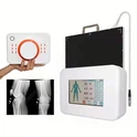
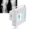
An X-ray machine is an essential tool in hospitals and clinics. It creates images of the body's internal structures using electromagnetic radiation. This technology is widely used in various medical fields.
An X-ray machine comprises several key components that work together. Its primary function is to capture images that help diagnose and monitor conditions. These machines offer insights into health issues without the need for invasive procedures.
Key features of an X-ray machine include:
A tube that emits X-rays
A detector to capture the image
A control panel for operation
X-ray machines are pivotal in medical imaging and diagnostics. They provide critical information on fractures, tumors, and infections, significantly aiding in patient care.

How Do X-Ray Machines Work?
X-ray machines operate using a fascinating principle. They emit X-rays, a form of electromagnetic radiation, which passes through the body. Different tissues absorb X-rays at varying rates, creating a detailed image.
When X-rays penetrate the body, dense materials like bones absorb more of them. This absorption makes bones appear white on the resulting image. Meanwhile, softer tissues allow more X-rays to pass through, so they appear in shades of gray.
The process involves several crucial steps:
Generating X-rays using a tube
Directing the X-rays toward the targeted body area
Capturing the image with a detector
Displaying the image for examination
The technology was discovered by Wilhelm Conrad Roentgen in 1895. Since then, X-ray machines have evolved, enabling clearer and faster imaging. They form the backbone of diagnostic imaging, assisting in the identification of fractures, infections, and foreign objects.
X-ray imaging is essential in healthcare, providing valuable insights. It guides treatment decisions, often being the first step in the diagnostic process. As such, understanding their operation is beneficial for both medical professionals and those curious about medical technology.

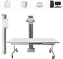
Key Components of an X-Ray Machine
An X-ray machine consists of several vital parts working in unison. Each component has a unique role in capturing high-quality images for medical diagnostics.
The core components include:
X-ray Tube: Produces X-rays aimed at the examination area.
Detector: Captures X-rays that pass through the body, forming an image.
Control Panel: Allows technicians to adjust settings and manage the imaging process.
The tube generates X-rays, which are focused on the specific body area of interest. This focusing is critical to obtaining accurate images. Meanwhile, the detector acts as a receptor, ensuring clarity and precision in imaging.
Modern machines enhance these traditional components with digital displays and advanced software. Such enhancements ensure improved image processing and reduced radiation exposure.

Applications in Diagnostic and Medical Imaging
X-ray machines have diverse applications in the realm of diagnostic imaging. Their versatility makes them indispensable in modern medicine.
In orthopedic medicine, X-rays are crucial for diagnosing bone fractures and joint injuries. They allow doctors to see the alignment of bones, aiding in precise treatment planning. Moreover, X-ray imaging is extensively used in dentistry to evaluate oral health. It helps in spotting cavities, tooth decay, and jawbone issues.
Beyond these, X-ray machines are applied in:
Cardiology: Detecting heart and lung conditions.
Mammography: Screening for breast cancer.
Emergency Medicine: Quickly assessing trauma injuries.
Each application harnesses the power of X-ray technology to provide accurate assessments. This is vital for effective medical interventions. As patient safety is a priority, technicians use the lowest radiation dose necessary.
The versatility of X-ray machines extends even further with the use of contrast agents. These enhance the visibility of soft tissues, providing clearer images in certain examinations. Their widespread use highlights their significance in healthcare.

Safety and Radiation Protection
Safety is a paramount concern with X-ray machines due to radiation exposure. Proper measures are essential to protect both patients and technicians.
Various safety protocols are in place to minimize risk. Technicians use lead aprons and thyroid shields to block radiation. These barriers significantly reduce exposure during X-ray procedures. Moreover, limiting the duration and frequency of X-ray usage further protects patients.
Key safety practices include:
Routine maintenance: Ensures machines function safely.
Patient positioning: Reduces unnecessary exposure.
Usage of the lowest effective dose: Balances safety and image quality.
Radiology staff undergo rigorous training to maintain these standards. This commitment to safety underscores the responsibility in using X-ray technology wisely. Focusing on protection helps ensure that diagnostic benefits do not compromise patient health.
Advances in X-Ray Technology
X-ray technology has evolved significantly over the years. These advancements have improved diagnostic accuracy while reducing radiation exposure. Modern innovations cater to diverse medical needs.
Recent developments include digital X-ray machines and portable devices. Digital systems offer quick image processing and better quality. This helps in more accurate diagnoses for healthcare professionals. Moreover, portable X-rays bring imaging to bedside, ideal for critical care settings.
Key technological advances include:
3D imaging: Provides detailed views of complex structures.
AI integration: Enhances image interpretation and diagnostics.
Lower radiation doses: Improves patient safety without compromising image clarity.

These breakthroughs have transformed how medical imaging operates today. They continue to drive innovations that benefit patient care across healthcare settings.
The Importance of X-Ray Machines in Healthcare
X-ray machines are vital in modern healthcare. They play a key role in diagnosing numerous conditions quickly and effectively. From identifying fractures to detecting tumors, these machines offer essential insights into a patient's health.
The benefits of X-ray machines in healthcare include:
Fast diagnosis: Enables prompt medical intervention.
Non-invasive examinations: Minimizes patient discomfort.
Guidance for further testing: Directs necessary follow-up procedures.
Their ability to provide immediate, visual information is unmatched. This technology ensures that medical professionals can make informed decisions quickly. Consequently, X-ray machines have become indispensable tools in hospitals and clinics globally.
Conclusion
X-ray machines revolutionize medical diagnostics, offering unparalleled insights into the human body. Their ability to deliver rapid results makes them indispensable in healthcare settings. Continuous advancements in technology aim to enhance image quality while minimizing radiation exposure.
Understanding the basics of X-ray machines highlights their vital role in improving patient outcomes. Their consistent evolution ensures they remain at the forefront of diagnostic imaging.