Understanding the Basics of Traditional X-Ray Machines
Traditional X-ray machines are a cornerstone of medical imaging. They have been used for over a century. These machines help diagnose a wide range of conditions.
X-ray machines work by sending X-ray beams through the body. This creates images of bones and tissues. The images are crucial for medical diagnosis.
Understanding how these machines work is important. It helps in appreciating their role in healthcare. It also highlights the need for safety measures.
The components of an X-ray machine are fascinating. They include the X-ray tube and image receptor. Each part plays a vital role in image creation.
X-ray technology has evolved significantly. Despite advancements, traditional machines remain essential. They are widely used in hospitals and clinics.
This article explores the basics of traditional X-ray machines. It covers their history, components, and applications. It also discusses safety and future trends.
What Is a Traditional X-Ray Machine?
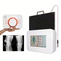
A traditional X-ray machine is a medical device used in diagnostic imaging. It utilizes X-ray beams to create images of the body's internal structures. X-ray machines are essential for identifying various health issues.
The primary function is to visualize bones and organs. This helps in detecting fractures, infections, and abnormalities. X-ray machines produce images that aid in diagnosis and treatment planning.
Key features of a traditional X-ray machine include:
X-ray tube
Control panel
Image receptor
Collimator for guiding the beam
These components work together to generate images. The X-ray tube emits X-rays, which pass through the body. The image receptor captures the resulting image for analysis.

The images produced are often black and white. Denser structures like bones appear white, while softer tissues appear darker. This contrast helps differentiate between various anatomical features, providing valuable clinical insights.
The History and Evolution of X-Ray Technology
X-ray technology has a fascinating history. Wilhelm Conrad Roentgen discovered X-rays in 1895. This groundbreaking discovery revolutionized medical diagnostics and imaging techniques.
The initial X-ray machines were simple and rudimentary. Over time, they evolved into sophisticated devices with enhanced capabilities. Key milestones in the evolution of X-ray machines include:
Introduction of digital systems
Improved image quality
Enhanced safety features
Development of portable devices

The integration of digital technology marked a significant advancement. It improved image quality and reduced radiation exposure. Portable X-ray machines further expanded access to remote and underserved areas, enhancing healthcare delivery.
As X-ray technology continues to evolve, it integrates with modern techniques like AI for better diagnostics. These continuous innovations ensure that X-ray imaging remains a cornerstone of medical practice, providing critical insights for patient care.
How Traditional X-Ray Machines Work
Traditional X-ray machines function by utilizing electromagnetic radiation. This process begins with generating X-rays from an X-ray tube within the device. The tube plays a pivotal role in producing the high-energy beams needed for imaging.
These X-ray beams penetrate the patient's body, passing through tissues and organs. Different tissues absorb X-rays to varying degrees, enabling the creation of distinct images. Denser structures like bones absorb more radiation, appearing white on the resulting images.
Key steps in the X-ray imaging process include:
Generating X-rays via the X-ray tube
Beams passing through the patient
Capturing images on receptors

The image receptor captures the X-ray image for further analysis. It can be either film-based or digital, depending on the system. Film-based systems have been largely replaced by digital receptors that provide clearer images and easier storage.
Overall, understanding the operation of traditional X-ray machines is crucial for healthcare professionals. This knowledge ensures accurate diagnostics while maintaining safety standards for both patients and operators.
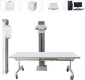
Key Components of a Traditional X-Ray Machine
Traditional X-ray machines comprise several essential components, each playing a vital role in the imaging process. Understanding these components helps ensure the effective use and maintenance of the equipment.
The main components include:
X-ray tube: Generates the X-rays needed for imaging.
Control panel: Allows technicians to adjust exposure settings.
Image receptor: Captures the X-ray image after beams pass through the patient.

The X-ray tube is the heart of the machine, producing beams by accelerating electrons. These electrons collide with a metal target, resulting in the emission of X-rays. This controlled process is crucial for safe and accurate imaging.
The control panel is where technicians set parameters like exposure time and intensity. These adjustments ensure that the X-ray beams are tailored to the needs of the patient and the specific diagnostic task.
Finally, the image receptor can either be a traditional film or a modern digital plate. Digital receptors have revolutionized medical imaging by providing enhanced image quality and easier management. Each component is integral to the overall function and success of traditional X-ray machines.
Common Applications in Medical Imaging
Traditional X-ray machines play an indispensable role in medical imaging. They are widely used across various healthcare settings due to their versatility and efficiency.
One of their primary uses is in the examination of bones. X-rays can easily detect fractures and monitor bone healing over time. This capability is crucial for orthopedic and emergency medicine.
Another common application is in dental imaging, where X-rays help in assessing teeth and jaw conditions. Dentists frequently rely on X-ray images to guide treatment decisions for cavities, impacted teeth, and more.

In addition to bones and teeth, traditional X-ray machines are also utilized for examining chest conditions. They help in diagnosing lung issues, such as infections, tumors, and fluid accumulation. The ability to diagnose various health conditions swiftly makes traditional X-ray machines vital tools in medical diagnostics.
Advantages of Traditional X-Ray Machines
Traditional X-ray machines offer several notable advantages, making them essential in healthcare. One key benefit is their cost-effectiveness, allowing widespread use in various settings.
These machines provide quick and non-invasive imaging. The speed and simplicity of the procedure make it ideal for emergency and routine exams.
Additionally, traditional X-ray machines are versatile. They cater to many diagnostic needs, from bones to soft tissues.
Cost-effective and accessible
Quick, non-invasive imaging
Versatile across diagnostic needs
Their continued use is testament to their reliability in providing vital diagnostic insights, contributing significantly to patient care.
Limitations and Challenges
Traditional X-ray machines, while effective, have some limitations. They rely on radiation, which poses health risks with frequent exposure.
Image quality can be another issue, especially when capturing soft tissues. Digital systems often provide better clarity and detail.
Operational constraints also arise. Large and stationary machines are hard to use in some medical settings.
Radiation exposure risks
Limited soft tissue detail
Operational and mobility constraints
These challenges make it important for healthcare providers to weigh the benefits and risks. Selecting the right imaging modality is crucial for accurate diagnosis and patient safety.
Safety Measures and Radiation Protection
Radiation safety is a top priority in the use of traditional X-ray machines. Both operators and patients must be shielded from unnecessary exposure.
Several protective measures are implemented. Lead aprons and thyroid shields are commonly used to safeguard sensitive areas. These reduce exposure and enhance patient safety.

Healthcare facilities adhere to the ALARA principle. This stands for "As Low As Reasonably Achievable." It guides the reduction of radiation doses without compromising image quality.
Use of lead aprons and shields
Adherence to ALARA principle
Monitoring radiation exposure levels
Continuous monitoring of exposure levels is critical. This ensures safety protocols remain effective and updated. By following stringent guidelines, the potential risks of radiation can be minimized significantly.
Maintenance, Regulation, and Training
Regular maintenance of traditional X-ray machines is essential. It ensures optimal performance and accurate diagnostic results. Scheduled inspections help in identifying and rectifying issues early.
Regulation is another critical aspect of using X-ray machines. Regulatory bodies set standards for equipment quality and safety. Compliance with these regulations is mandatory in healthcare facilities.
Training is crucial for operators. Radiology technicians need to understand equipment operation and safety protocols. Proper training minimizes mistakes and maximizes patient safety.
Regular maintenance schedules
Compliance with regulatory standards
Comprehensive operator training programs
Adhering to these practices helps maintain the integrity of X-ray systems. Proper education and compliance ensure safe and effective medical imaging.
The Role of Traditional X-Ray Machines in Modern Healthcare
Traditional X-ray machines play an essential role in medical imaging. They are often the first step in diagnostic evaluations. Quick and accurate imaging aids in effective patient care.
In emergency settings, X-ray machines provide rapid assessments. They help in diagnosing fractures and other urgent conditions. This efficiency is critical for timely treatment.
X-ray machines also support routine exams. They are valuable in dental and orthopedic practices. The versatility and accessibility of X-ray technology make it indispensable.
Initial diagnostic assessments
Emergency and trauma diagnosis
Routine examination support
Traditional X-ray machines continue to be a cornerstone in healthcare. Their importance in diagnosing and managing health issues is unmatched.
Future Trends and Innovations in X-Ray Technology
Innovations in X-ray technology focus on improving image quality. Reduced radiation exposure is also a significant goal. Digital advancements streamline the imaging process.
Artificial intelligence is making strides in this field. It aids in image analysis and interpretation. This integration allows for faster, more accurate diagnostics.
Emerging Trends
Advanced imaging techniques
AI integration in diagnostics
Enhanced safety measures

These trends aim to enhance patient care and safety. The future of X-ray technology promises more efficient solutions. Continuous research fuels these exciting developments.
Conclusion: The Enduring Value of Traditional X-Ray Machines
Traditional X-ray machines remain vital in medical imaging. Despite technological advancements, their role is irreplaceable in diagnostics.
These machines offer quick and reliable results. Their enduring presence in healthcare ensures accessibility and effective patient care. The future holds even more advancements, building upon these steadfast foundations.