Understanding the Basics of Traditional X-Ray Machines
Traditional x-ray machines are vital in medical diagnostics. They provide clear images of the body's internal structures.
These machines have been a cornerstone in healthcare for over a century. They help detect fractures, infections, and tumors.
X-ray machines work by emitting controlled radiation. This radiation passes through the body to create images on film or digital sensors.
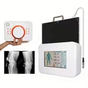
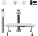
The main components include the x-ray tube, control panel, and image receptor. Each part plays a crucial role in image production.
Traditional x-ray machines are often more cost-effective than advanced imaging technologies like CT or MRI. This makes them accessible to many healthcare facilities.
Safety is paramount in radiology. X-ray machines must adhere to strict standards to minimize radiation exposure.
Regular maintenance and calibration are essential. They ensure accurate and safe diagnostic results.
Understanding x-ray technology is important for healthcare professionals. It enhances their ability to provide effective patient care.

What Is a Traditional X-Ray Machine?
A traditional x-ray machine is a standard piece of radiology equipment. It has been used in medicine for over a century.
These machines utilize x-rays, a form of electromagnetic radiation. They produce images of bones and soft tissues.
X-ray machines are integral to medical imaging. They help physicians diagnose a variety of conditions.
Core Features of Traditional X-Ray Machines
X-Ray Tube: Generates x-rays using high voltage.
Control Panel: Allows technicians to adjust settings.
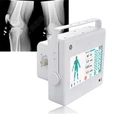
Image Receptor: Captures the x-ray images.
Traditional x-ray machines are distinct from digital models. They often use physical film to capture images.
The development of digital systems has changed radiography. However, traditional machines remain in use due to their cost-effectiveness and reliability.
These machines are favored in many settings. They are especially prevalent in hospitals, clinics, and dental offices.
X-ray technology began with Wilhelm Conrad Roentgen in the 19th century. His discovery opened a new frontier in medical diagnosis.
Understanding traditional x-ray machines is key for medical professionals. This knowledge ensures effective use in patient care.

How Do Traditional X-Ray Machines Work?
Traditional x-ray machines function through precise mechanisms. They produce x-rays by releasing radiation through an x-ray tube.
The tube requires high voltage to work. This generates x-rays that can pass through the body.
As x-rays travel, they encounter different tissues. Each tissue absorbs x-rays differently due to varying densities.
The Process of Image Formation
X-Ray Generation: High voltage powers the x-ray tube.
X-Ray Emission: X-rays are directed towards the patient.
Tissue Interaction: X-rays pass through or are absorbed by tissues.
Image Capture: Image receptors or films record the x-rays that pass through.
The resulting image displays shadows and contrasts. Bones appear white while soft tissues show as shades of gray.
This imaging process allows healthcare providers to detect anomalies. They can spot fractures, detect infections, and monitor conditions.
A crucial aspect is the control panel. It allows radiologic technologists to adjust the radiation dose and exposure time.
These adjustments optimize image quality while ensuring patient safety. Balancing these factors is vital for effective diagnostics.
Safety is paramount when using x-ray machines. Operators follow strict guidelines to minimize radiation exposure.

Key Components of Traditional X-Ray Machines
Traditional x-ray machines consist of several essential parts. Each component plays a critical role in image creation.
The x-ray tube is the heart of the machine. It generates the x-rays needed for imaging.
Additionally, the control panel is where operators adjust the settings. This includes the x-ray dose and exposure time.
Another vital component is the image receptor. It captures the image formed by x-rays passing through the body.
To sum it up, key components include:
X-Ray Tube: Generates x-rays.
Control Panel: Adjusts exposure settings.
Image Receptor: Captures the resulting image.
Patient Table: Supports patient during the procedure.
Collimator: Focuses x-rays on the target area.
The patient table ensures stability during imaging. It supports varied patient positions for optimal results.
Collimators help to direct x-rays precisely. They reduce scatter radiation, improving image clarity and safety.
Understanding these components aids in grasping the machine's functionality. It highlights the complexity and precision involved in medical imaging.
Regular maintenance is crucial for efficient operation. It ensures that each component performs correctly, yielding accurate diagnostic images.

The Role of Diagnostic X-Rays in Medicine
Diagnostic x-rays are invaluable tools in modern medicine. They enable doctors to look inside the body non-invasively.
X-rays reveal the internal structure of bones and organs. This helps in diagnosing conditions without the need for surgery.
They are particularly useful in detecting fractures and bone disorders. X-rays also uncover hidden infections or tumors.
In emergency rooms, x-rays provide quick diagnostic support. They help doctors make critical decisions rapidly.
Some common medical applications of diagnostic x-rays include:
Fracture Detection: Identifying broken bones.
Chest X-Rays: Evaluating lung conditions.
Dental X-Rays: Assessing oral health.
Mammography: Screening for breast cancer.
Fluoroscopy: Real-time imaging for procedures.
Radiography is critical in treatment planning. It guides surgeries and monitors healing processes.
The accessibility and speed of x-rays make them indispensable. This is particularly true in urgent medical scenarios.
Understanding the role of diagnostic x-rays aids healthcare professionals. It ensures that patients receive timely and accurate care.
Overall, diagnostic x-rays continue to be a cornerstone of clinical practice. Their role in improving patient outcomes is undeniable.

Common Applications and Uses
Traditional x-ray machines serve diverse functions in healthcare. They are widely employed across various medical fields.
One primary use is in orthopedics. X-rays detect fractures and monitor bone healing effectively.
These machines also play a crucial role in dentistry. Dentists use them to examine teeth, gums, and jaw alignment.
Chest x-rays are another important application. They assess lung conditions and monitor heart size.
Other common uses of traditional x-ray machines include:
Abdominal Scans: Detecting blockages or foreign objects.
Skeletal X-Rays: Diagnosing bone-related ailments.
Trauma Cases: Quickly identifying internal injuries.
Veterinary Medicine: Assisting in animal care diagnostics.
In emergency medicine, speed matters. X-rays offer fast imaging solutions in critical situations.
Furthermore, these machines are cost-effective. They remain a practical choice for many healthcare providers.
Traditional x-ray machines are essential tools. They provide valuable insights into patient health swiftly and accurately.

Safety Measures and Radiation Protection
Safety is paramount when using traditional x-ray machines. These devices emit radiation, necessitating stringent protective measures.
Healthcare professionals prioritize minimizing exposure. This ensures the safety of patients and staff alike.
Protective gear like lead aprons is common. They shield sensitive areas from unnecessary radiation.
Operators take several measures to enhance safety, including:
Limiting Exposure Time: Reduces the duration of radiation exposure.
Using Shielding Devices: Such as lead barriers and portable shields.
Maintaining Safe Distances: Keeping a safe distance from active x-ray machines.
Regular machine maintenance is crucial too. It ensures devices function correctly and safely.
Calibrating x-ray machines helps maintain optimal performance. This step is vital for accurate diagnostics.
Adhering to safety standards is mandatory. Regulatory bodies set guidelines to protect all involved.
Patient consent is also important. Informing patients about benefits and risks helps in decision-making.
Advancements in technology continue to improve safety. New features aim to further reduce radiation exposure.
Implementing these precautions helps maintain a safe diagnostic environment. This is essential for effective healthcare delivery.

Traditional X-Ray Machine Cost and Maintenance
Investing in traditional x-ray machines involves understanding their costs. Prices vary depending on several factors, including model and manufacturer.
While more affordable than advanced imaging devices, costs can still be significant. It's important to consider both initial purchase and ongoing expenses.
Key factors influencing cost include:
Brand and Model: Well-known brands may cost more, offering advanced features.
Features and Specifications: Machines with more capabilities tend to be pricier.
New vs. Refurbished: Refurbished units are often more budget-friendly.
Maintenance is crucial for optimal performance and safety. Regular servicing ensures machines work accurately and last longer.
Routine checks and calibrations are necessary. These prevent errors and improve diagnostic accuracy.
Budgeting for maintenance helps avoid unexpected expenses. It's important for healthcare facilities to plan accordingly.
Training staff in basic troubleshooting can save costs. It reduces the need for frequent professional repairs.
Selecting the right machine involves weighing cost against benefits. Consider long-term needs and potential returns on investment.
Proper upkeep prolongs machine lifespan, ensuring reliable diagnostic results. This supports efficient healthcare service delivery.

Traditional vs. Digital X-Ray Machines
Choosing between traditional and digital x-ray machines depends on various factors. Both types have unique advantages and limitations.
Traditional x-ray machines have been in use for over a century. They are reliable and familiar to many healthcare professionals.
Digital x-ray machines offer advanced technology. They provide better image quality and quicker results.
Key differences include:
Image Quality: Digital offers higher resolution images.
Speed: Digital machines process images faster.
Cost: Traditional machines are usually more cost-effective.
Maintenance: Digital systems require specific technical expertise.
Despite advancements, traditional machines remain popular. They deliver consistent results, especially in less complex diagnostics.
For budget-conscious facilities, traditional units are appealing. They cost less initially and often require simpler upkeep.
However, digital systems can streamline workflows. They reduce the need for physical film and associated storage.
Facilities must weigh needs, budget, and patient volume. This ensures the best choice for their specific diagnostic requirements.
Future trends may see more integration of digital technology. However, traditional machines continue to be vital assets.

Advances in Radiology Equipment and Future Trends
Radiology equipment continues to evolve, offering improved imaging capabilities. The focus is on enhancing accuracy and safety for patients and operators.
Innovations include developments in digital x-ray technology. This advancement leads to superior image quality with reduced radiation levels.
Recent improvements also streamline data management. Systems like PACS enable efficient storage and retrieval of diagnostic images.
Key future trends to watch for:
AI Integration: Assisting radiologists in image analysis.
Portable Devices: Lightweight, easy-to-transport x-ray units.
Enhanced Safety: New materials and designs minimizing radiation exposure.
Faster Processing: Rapid imaging aiding quick diagnostics.
These trends demonstrate the dynamic nature of radiology. As technology advances, healthcare providers can deliver better patient care.
The use of AI in radiology is particularly promising. It can improve diagnostic accuracy and reduce human error.
Despite the push for digital solutions, traditional machines hold their ground. They continue to serve essential roles in various medical settings.

Training and Certification for X-Ray Technologists
Operating x-ray machines requires specialized skills. Technologists must undergo rigorous training to ensure competence and safety.
Certification is crucial for these professionals. It verifies their ability to handle complex radiology equipment effectively.
Key training components include:
Radiation Safety: Understanding safe exposure levels.
Equipment Operation: Handling both traditional and digital machines.
Image Analysis: Developing skills to assist in diagnostics.
Patient Care: Ensuring comfort and effective communication.
Continuing education is also essential. It helps technologists stay updated on the latest advancements and techniques.
Certification and ongoing training ensure high-quality diagnostic care. They play a crucial role in maintaining patient and operator safety.
Conclusion: The Enduring Value of Traditional X-Ray Machines
Traditional x-ray machines continue to hold significant value in the medical field. Their cost-effectiveness, reliability, and rapid imaging capabilities make them a staple in diagnostics.
Despite advances in digital technology, traditional machines remain prevalent. They offer dependable service and vital insights into patient health. Their ongoing use underscores their lasting importance in radiology and healthcare.