A Comprehensive Overview of Digital X-Ray Technology and Its Evolution in Medical Imaging
The development and evolution of X-ray technology have significantly impacted the medical field, revolutionizing the way diseases are diagnosed and treated. The journey began with the groundbreaking discovery of X-rays by German physicist Wilhelm Roentgen on November 8, 1895. This discovery marked the dawn of radiology, a new medical discipline that has since become an integral part of modern healthcare. Over the past century, X-ray technology has undergone remarkable advancements, culminating in the widespread adoption of digital X-ray equipment, which now dominates the medical imaging landscape.
The Evolution of X-Ray Technology
Since its inception, X-ray technology has steadily evolved to meet the growing demands of the medical field. In the early 20th century, radiology emerged as a distinct medical discipline, enabling physicians to diagnose and treat a wide range of diseases using X-ray imaging. This new era of medical diagnostics revolutionized healthcare, allowing for non-invasive examination of the human body. As technology progressed, so did the sophistication of X-ray equipment, leading to the development of digital X-ray systems that offer superior image quality, reduced radiation exposure, and faster processing times.
Digital X-ray technology represents a significant leap forward from traditional analog systems. The transition to digital imaging has brought about numerous benefits, including enhanced image clarity, improved diagnostic accuracy, and more efficient workflows. Today, digital X-ray machines are ubiquitous in hospitals, clinics, and diagnostic centers worldwide, playing a crucial role in the early detection and management of various medical conditions.
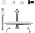
Components of Digital X-Ray Diagnostic Equipment
Digital X-ray diagnostic equipment is composed of several key components, each playing a vital role in the imaging process. These components include the X-ray generator, the X-ray imaging device, and various accessory devices that support the overall functionality of the system.
X-Ray Generator: The X-ray generator is responsible for producing X-rays and controlling their output. It typically consists of an X-ray source, a high-voltage generator, and control devices. The generator ensures that the appropriate amount of radiation is emitted, which is crucial for capturing clear and accurate images.
X-Ray Imaging Device: The imaging device is where the magic of digital X-ray technology truly shines. It comprises a flat-panel detector, a computer system, and image processing software. The flat-panel detector converts the X-ray signal into a digital signal, which is then processed by the computer to produce a digital X-ray image. Advanced image processing software enhances these images, making it easier for radiologists to detect and diagnose various conditions. This includes automatic adjustments to contrast and brightness, noise reduction, and even specialized imaging techniques like long limb stitching and energy subtraction.
Accessory Devices: Accessory devices include all the supporting equipment necessary for the operation of digital X-ray systems. This encompasses diagnostic beds, support structures, suspension devices, brake systems, and beam control devices. These components work together to ensure that the X-ray system operates smoothly and efficiently, providing accurate and reliable results.
The Advantages of Digital Radiography
Digital radiography (DR) is a modern X-ray imaging technique that directly captures digital images under the control of a computer. Unlike traditional optical imaging, digital radiography offers several key advantages:
Enhanced Image Quality: Digital radiography produces images with superior clarity and detail, making it easier to detect even subtle abnormalities. The high-definition images generated by DR systems allow for more accurate diagnoses, particularly in complex cases where traditional X-ray methods may fall short.
Lower Radiation Dose: One of the most significant benefits of digital radiography is the reduced radiation exposure for patients. DR systems are designed to use lower doses of radiation while still producing high-quality images, which is particularly important in minimizing the risks associated with repeated imaging.
Faster Processing and Results: The speed of digital radiography is another major advantage. Images are available within seconds of exposure, allowing radiologists to quickly assess the results and make informed decisions. This rapid turnaround is especially beneficial in emergency situations where time is of the essence.
Versatility in Imaging Applications: Digital radiography is used in a wide range of medical applications, from chest X-rays for respiratory conditions to musculoskeletal imaging for bone and joint issues. It is also commonly employed in abdominal imaging, urinary system diagnostics, and breast cancer screening through mammography.
Applications of Digital Radiography in Healthcare
Digital X-ray technology has become a cornerstone of medical imaging, with applications across various medical specialties. Here are some of the most common uses of digital radiography:
Respiratory System: Chest X-rays are the standard imaging method for diagnosing respiratory diseases. Digital radiography is particularly effective in detecting lung conditions such as pneumonia, tuberculosis, and lung cancer. The high contrast and detailed images provided by DR systems enable early detection and more accurate assessments of pulmonary diseases.
Musculoskeletal System: Digital radiography is the preferred imaging technique for evaluating bone and joint conditions. It is widely used to diagnose fractures, arthritis, and other musculoskeletal disorders. The ability to capture clear images of both bone and soft tissue structures makes DR an invaluable tool in orthopedic diagnostics.
Abdominal Imaging: In cases of acute abdominal conditions, such as gastrointestinal perforations or obstructions, digital radiography provides critical diagnostic information. The fast processing time and high image quality make it an essential tool in emergency medical care.
Urinary System: Digital radiography is used to identify urinary stones and assess renal function. Intravenous nephrography, for example, utilizes X-ray imaging to visualize the renal pelvis and ureters, helping to diagnose conditions such as kidney stones and urinary tract obstructions.
Breast Cancer Screening: Mammography, a specialized form of digital radiography, is a crucial tool in the early detection of breast cancer. Modern mammography systems, such as digital breast tomosynthesis (DBT), offer enhanced imaging capabilities that improve the detection of small tumors and reduce the need for follow-up biopsies.
The Future of Digital X-Ray Technology
As technology continues to advance, digital X-ray systems are expected to become even more sophisticated. Innovations such as artificial intelligence (AI) and machine learning are already being integrated into imaging software, enabling automated image analysis and more accurate diagnoses. Additionally, the development of portable and mobile X-ray units is expanding access to high-quality imaging in remote and underserved areas.
In conclusion, digital X-ray technology has transformed the field of medical imaging, offering numerous benefits over traditional methods. From enhanced image quality and lower radiation exposure to faster processing times and versatile applications, digital radiography is an essential tool in modern healthcare. As technology continues to evolve, the future of digital X-ray imaging looks brighter than ever, with the potential to further improve patient outcomes and revolutionize the way we diagnose and treat medical conditions.